Bladder Tumors
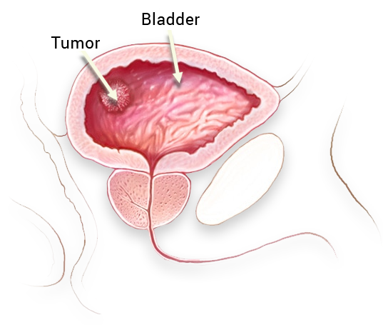
The bladder is a hollow muscular organ that collects and disposes of the urine that is filtered by the kidneys. Bladder cancer is most frequently caused by the cells (transitional epithelium cells) lining the inside of the bladder.
The bladder is a hollow muscular organ that collects and disposes of the urine that is filtered by the kidneys. Bladder cancer is most frequently caused by the cells (transitional epithelium cells) lining the inside of the bladder.
There are mainly 3 types of cancer (malign tumors) that might develop in the bladder.
- Transitional cell carcinoma (Urothelial carcinoma):
In this type of cancer, the cancer is caused by the cells lining the inside of the bladder, which are called epithelium cells. It is the most common type of bladder cancer. - Squamous cell carcinoma:
The cancer is caused by the squamous cells developing in the bladder as a result of long-term exposure of the bladder to infection and irritation. - Adenocarcinoma:
This type of cancer is caused by the glandular (secretory) cells in the bladder. These secretory glandular cells line the inside of the bladder and produce mucus.
Bladder cancer is the fourth most common type of cancer in the United States and the fifth in Europe. According to the data from the NCI (National Cancer Institute), 68,810 people were diagnosed with bladder cancer and 14,100 deaths occurred from bladder cancer in 2008 in the United States. Bladder tumors are 3 times more common among men than women. Prostate cancer is the fourth most common cancer among men, following lung and colon cancers. They make up 7% of the cancer cases among men. It is the ninth most common cancer among women, making up 2.5% of all cancer cases.
Bladder cancer can occur at any age, including in childhood. However, it is generally seen among middle-aged and old-aged people. The prevalence rises with increasing age.
It is reported that inactivation of various tumor suppressor genes has a role in development of bladder cancer. Today, the most prominent tumor suppressor genes are TP53 and the cell cycle inhibitors are RB, P21, P27 and P16, which are shown to be associated with development of bladder cancer.
Exposure to environmental carcinogens is also significant in terms of development of bladder cancer. Bladder cancer is more common among chimney sweepers and people working in plastic and rubber industries. Additionally, bladder cancer rate is 4 times higher among smokers than non-smokers.
Signs and Symptoms
The most common sign of bladder cancer is painless streaks of blood in urine. Painless and intermittent streaks of blood in urine occurs in nearly 85% of the patients. Symptoms like frequent urination, burning while urinating and difficulty urinating might be the first signs of a bladder tumor. Clots might sometimes accompany the blood in the urine. In addition, symptoms might include pain in the lower abdomen or lumbar region.
Diagnosis
Urine tests play an important part in diagnosis. A complete urinalysis showing blood cells (erythrocytes) in urine should suggest presence of a tumor.
Urine Cytology
It is a method involving analysis of urine by a pathologist for detection of cancer cells. Urine cytology is a method that has low sensitivity in case of low-grade tumors and has a maximum sensitivity of 80% in case of high-grade tumors.
Other Urine Analyses
Today there are certain tests available for use in diagnosis. BTA stat and NMP22 are among the tests that are also used in our country. However, they have low sensitivity for small tumors. Others include ImmunoCyst and UroVision DNA FISH tests. Whereas ImmunoCyst probably has the highest sensitivity for small and low-grade tumors, UroVision DNA FISH has the highest specificity.
Cystoscopy
This is the easiest and surest method of establishing a diagnosis in case of a suspected bladder tumor. An optical device is advanced through the urethra and the inside of the bladder can be viewed with 10 times magnified images. Flexible cytoscopy is a simple yet very valuable diagnosis method that can be performed even under local anesthesia.
Imaging Methods
The simplest imaging method in diagnosis of bladder tumors is abdominal ultrasonography. A tumor that has reached a certain size can be viewed with ultrasound. Other methods include Computed Tomography (CT), Magnetic Resonance Imaging (MRI) and Intravenous Pyelogram (IVP). Additionally, Positron Emission Tomography (PET) is very useful in showing the degree of spread of this disease.
Staging
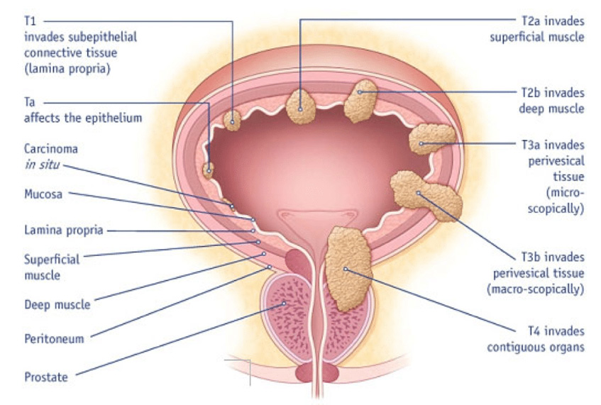
The wall of the bladder wall consists of 3 main layers:
- Mucosa
- Lamina propria
- Muscularis propria (muscular layer).

Staging
Ta tumors only involve the mucosa. T1 tumors, on the other hand, have spread to the lamina propria. The tumor has spread to the superficial muscular layer in T2 stage and the deep muscular layer in T3 stage. Tumors called carcinoma in situ (Tis) means the tumor is located only in the mucosa but could be dangerous for the patient if they do not receive the right treatment on time.
Aside from the clinical stating described above, there is also a microscopic staging that determines the behavior of the tumor. Whereas low-grade tumors are expected to display a better behavior, high-grade tumors are expected to progress more aggressively.
In case of a suspected bladder tumor, the tumor should be resected by entering through the urethra under general/lumbar anesthesia (Transurethral Resection, TUR-P). The tumor tissues resected by means of an electrocautery device should be sent for pathological analysis. In some cases, biopsy samples should be retrieved also from the normal-looking parts of the bladder.
In case of superficial tumors (Ta, T1), TUR-P may offer a permanent recovery. However, risk of recurrence is higher in the presence of multiple tumors or a tumor that has a diameter greater than 4 cm; in which case, the inside of the bladder should be irrigated with drug solutions like Mitomycin or Immun BCG once a week for 6-8 weeks.
If the tumor involves the muscular layers (T2, T3), the best treatment method is the total removal of the bladder (Radical Cystectomy), particularly in young patients who are in good overall health, and colon bladder placement after the cystectomy. For suitable patients, orthotopic bladder (colon bladder connected to the urethra) is the best method in terms of comfortableness after the surgery. For patients who are not candidate for this method, ileal loop (the method which allows the patient to carry a urine pouch on their abdomen) should be applied. Today, these procedures are performed through laparoscopic and even robotic means.
Follow-up
A patient with a bladder tumor always has the risk of recurrence. Therefore, patients should be followed up periodically, at the intervals to be recommended by the physician. Accordingly, urine tests and control cystoscopy should be performed on a regular basis. Patients who have been diagnosed with a superficial tumor might have a low risk of total removal of their bladders at some point in their lives. This risk is lower in case of low-grade tumors while higher in high-grade tumors. In case of high-grade tumors, there is a little risk of tumor developing in the upper urinary system – kidney pelvis and the urethra, where the urine is collected and sent. Therefore, kidneys should also be examined once every 2 years.

Bladder Tumors
The bladder is a hollow muscular organ that collects and disposes of the urine that is filtered by the kidneys. Bladder cancer is most frequently caused by the cells (transitional epithelium cells) lining the inside of the bladder.
Book an Appointment with Dr. Fatih Atug
Cantact Dr. Fatih Atug
Fatih Atug, M.D.
Urologist and Robotic Surgery Specialist
hidden +90 212 234 5958
hidden +90 532 234 5504
hidden info@fatihatugmd.com
hidden Harbiye Mahallesi, Maçka Caddesi,
Bahriye Apt. No:13 D: 3
Şişli / İSTANBUL
 |
 |
 |


